



Programme 01200 / 200 ADMITTED
MBBS
Undergraduate · Phase I Curriculum
Seats permitted200
Structured exposure to gross anatomy, histology, and embryology with integrated clinical correlates from Day 1.
Established in 1999, the Department of Anatomy is dedicated to excellence in human anatomical education and research, integrating traditional dissection with advanced AR-VR technology and pioneering plastination techniques.
The Department of Anatomy at AVMCH is dedicated to providing high-quality training in all branches of human anatomy. We adopt a student-centric approach, utilizing audiovisual-assisted lectures, small group discussions, and integrated teaching to build the foundation of medical excellence.
200 MBBS seats / yr
3 PG seats / yr
8 PhD seats permitted
Anatomy at AVMCH blends traditional dissection with immersive AR-VR technology and pioneering plastination to provide a world-class structural education.
The Department of Anatomy is committed to providing high-quality education and training in all branches of human anatomy for undergraduate and postgraduate students, in accordance with NMC guidelines. The department adopts a combination of traditional teaching methods and modern, student-centric approaches, including audiovisual-assisted lectures, small group discussions, and integrated teaching. It fulfills all essential requirements for medical education and contributes to continuous quality improvement in teaching, learning, and research.
Our postgraduate and doctoral programs (MS/MD and PhD) offer specialized training in Clinical and Applied Anatomy, Neurosciences, and Cytogenetics. The department features a dedicated research laboratory and specialized units such as the AR-VR Lab, Plastination Unit, and Anthropometric Lab. We maintain an active voluntary body donation program ensuring a consistent supply of cadavers for anatomical training and advanced surgical skill workshops.
The department provides advanced infrastructure for learning, including one of Puducherry's finest Anatomy Museums and well-equipped histology laboratories. Our teaching-learning facilities provide access to high-quality histological slides, clinically relevant radiographs, and CT scans. Specialized units like the Plastination Unit create durable, odorless specimens for long-term study, while the AR-VR Lab offers immersive 3D visualization of human structures.
The department maintains a well-equipped dissection hall, histology labs, and a state-of-the-art AR-VR lab for immersive learning.
Faculty and postgraduate students actively engage in clinical and applied anatomy research, supported by our voluntary body donation program.
Research Focus
Research Focus · Clinical Variations, Neuroanatomy & Cytogenetics
Outreach
Outreach · Body Donation Program · AR-VR Demos
Academic Programmes
Undergraduate · Phase I Curriculum
Structured exposure to gross anatomy, histology, and embryology with integrated clinical correlates from Day 1.
Postgraduate · 3-year specialty
Advanced training in cadaveric dissection, neuroanatomy, molecular biology, and research methodology.
Doctoral · Research programme
Doctoral research across clinical and applied anatomy, cytogenetics, and histology, supported by the department's research laboratory and plastination unit.
The department is led by Dr. T. Rajan, a recipient of the Life Time Education Achievement Award and an expert in clinical anatomy.
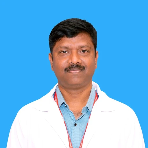
Professor & Head · MD, PhD Anatomy
An expert in plastination and clinical anatomy, Dr. T. Rajan leads the department's modernization through AR-VR and advanced preservation techniques. He has multiple publications in high-impact journals and is a recognized leader in basic medical sciences.
Capacity for 300 students with 30 large and 5 small cadaver tables and advanced AV-assisted demonstration aids.
Recognized as one of the best in Puducherry, featuring 300+ wet/dry specimens and embryology models.
Immersive 3D environment for visualizing human anatomy and performing simulated dissections.
Revolutionary technique creating odorless, durable specimens for clinical and research study.
Equipped for histological, cytogenetic, and anthropometric research with active faculty and PG involvement.
Specialized Embryology and Anthropometric labs for studying developmental and physical human variations.
5 qualified professionals committed to clinical excellence.
Anatomical variations and their implications in surgery and diagnostic imaging (CT/MRI).
Structural organization of the nervous system and functional correlations with clinical disorders.
Light microscopic studies of tissues, tissue response to physiological and pathological change, and advanced histological and staining techniques.
Chromosomal abnormalities and congenital disorders, karyotyping and genetic screening, and the molecular basis of structural and developmental anomalies.
Cadaver-based training and skill development, the anatomical basis of surgical approaches, and preservation techniques and body donation awareness.
Population-based morphometric studies, age, sex and stature estimation, and applications in forensic identification.
Normal and abnormal human development.
Innovative teaching–learning methods, digital tools, simulation and e-learning in anatomy education, and assessment under competency-based medical education (CBME).
The department focuses on Clinical and Applied Anatomy, Neurosciences, and Cytogenetics with active intramural and extramural funding.
Research Snapshot
10 projects since 2023 8 thrust areas
Intramural & extramural support
| # | Project | PI | Funding | Focus | Status |
|---|---|---|---|---|---|
| 01 | Anatomical variations of the extensor tendons over the dorsum of the hand | Dr. T. Rajan | Self-Funded | Clinical Anatomy | Ongoing |
| 02 | Interstitium is an Organ – Histochemical study | Mr. K. Sridhar | Extramural | Histology | Completed |
| 03 | Dermatoglyphics and its importance in precancerous and cancerous lesion | Dr. T. Rajan | Self-Funded | Clinical Anatomy | Completed |
| 04 | Characterization of oral cancer cell line Hep2: Targeted EGFR modulation | Dr. Hemavardhini R | Intramural | Molecular Anatomy | Ongoing |
| 05 | Effects of Allium Sativum on PCOS in female albino rats | Mrs. G. Sowmiya | Self-Funded | Histology | Ongoing |
| 06 | Morphology and morphometrical analysis of the anterior cruciate ligament in the adult human cadaveric knee | Dr. Jeffi Jerome (Postgraduate) | Nil | Cadaveric Anatomy | Completed |
| 07 | Neck-shaft angle and torsion angle of the adult human femur — an osteological study | Dr. Tokpam Rolinda (Postgraduate) | Nil | Osteology | Completed |
| 08 | Morphological and morphometric study of adult human dry lumbar vertebrae | Dr. Vimalananth J (Postgraduate) | Nil | Osteology | Completed |
| 09 | Morphological and morphometric evaluation of the sacral hiatus in dry bone and 3D reconstructed CT images | Dr. Shree Ranjany (Postgraduate) | Nil | Applied Anatomy | Ongoing |
| 10 | Histomorphological, biochemical and molecular analysis of short- and long-term stress-induced kidney of Swiss albino male mice | Dr. Leelavathy (Postgraduate) | Nil | Histology | Ongoing |
Int J Life Sci Pharma Res, 2023
Vinothini G, Elavarasi E, et al. (including Dr. T. Rajan)
Int J Life Sci Pharma Res, 2023
Prithiviraj N, Vinothini G, et al. (Corresponding: Dr. T. Rajan)
Clin of Diagn Res, 2023
Sudagar M, Bavithra B, Prithiviraj N
Journal of Medical Sciences, Vol 2, Issue 12
Sridhar K, Dr. T. Rajan, et al.
BMC Infectious Disease, 11.205, 2011
Padmanaban S Suresh, Venkatesh Thejaswini, Thangarasu Rajan
Journal of Infection, 2012
Padmanaban S. Suresh, Thangarasu Rajan, Venkatesh Thejaswini, Rajagopal Rajeshkannan
Endocrine Journal, 58(4), 223–235, 2011
Padmanaban S. Suresh, Thangarasu Rajan, Rie Tsutsumi
Gene, 511, 353–357, 2012
Padmanaban S. Suresh, Thejaswini Venkatesh, Thangarasu Rajan
Androl Gynecol: Curr Res, 1:3, 2013
Padmanaban S Suresh, Thejaswini Venkatesh, Thangarasu Rajan, Rie Tsutsumi
National Journal of Clinical Anatomy, 2(1), 31–34, 2013
Thangarasu Rajan, Atul M Bage
Futuristic Trends in Medical Sciences, Vol 3 (Book Chapter) · e-ISBN 978-93-6252-225-2
Dr. T. Rajan
300 Capacity
30 large and 5 small cadaver tables, an ante-room with lockers and washbasins, and AV aids with battery backup and LCD projection for specimen demonstration.
75 Capacity
Equipped with computer-assisted microscopes and TV demos for high-quality slide study.
Best in Class
Features 300+ wet/dry specimens, rotational X-ray view boxes, and embryology models.
Immersive
Uses 3D technology for interactive visualization and spatial exploration of human structures.
Specimen Preservation
Durable, odorless anatomical specimens created using advanced polymer replacement techniques.
Proportion Study
Used for human body dimension measurements for forensic and clinical growth assessments.
Philosophy
Anatomy · AVMCH
“Structural excellence in medical education — blending traditional dissection with immersive digital exploration.
Department of Anatomy, AVMCH & Hospital
Dr. T. Rajan
National & International Compendium, New Delhi, 2023
Dr. T. Rajan
Association of Anatomists, Chennai
Jodhpur
3rd National Annual Conference of Society of Medical Anatomists, 2026
15
Dec 2023
International Conference · Participants – 842 TMC – 6 credit hours
Organizing Chairman-Dr S.Ilankathir & Dr M.Gopinath Organizing Secretary – Dr M.Manju
04
Dec 2024
International e-conference · The conference had 542 registrations and 172 abstracts (141 papers for oral and 31 for young Research Scholar Award presentations in Faculty, PhD, Postgraduate and Undergraduate categories). E-Posters competition was also held for undergraduates, in which we had 5 entries. There were 13 eminent Resource persons (5 International and 8 National speakers) from UK, USA, Malaysia, and various parts of our country.
Dr S.Ilankathir, Organizing Secretary, Dr. M Manju, Organising Chairperson.
20
Jun 2025
CME on “Surgical Rehabilitation of the Deaf” · Total number of participants were 157 including UG, PG and faculties. Speaker: Dr. Sivaraman G, Additional Professor & Head, Department of ENT, JIPMER
Dr. Rajan T, Vice-Dean (Admin & Events), AVMCH Dr. S Ilankathir, Professor & Head, Department of Anatomy, AVMCH.
04
Jul 2025
Guest lecture titled "Peritoneum & Its Clinical Significance" · Total number of participants: 161 As part of the 25th Silver Jubilee Celebrations of Vinayaka Mission’s Puducherry CampusDr. Gladwin, JIPMER
Organized by: Department of Anatomy, AVMCH, Puducherry
03
Aug 2025
Workshop · Total Participants: 45 delegates. The workshop was met with enthusiastic participation and positive feedback. Live ultrasound-guided scanning Demonstrations of advanced procedures, such as nerve denervation, regenerative medicine approaches and cancer pain interventions, fostering confidence in performing these techniques safely and effectively in clinical settings.
Organising Secretary: - Dr. Rajan T, Vice-Dean (Admin & Events), AVMCH
25
Sep 2026
Workshop · Cadaveric Nerve Block Workshop in the Anatomy Dissection Hall, conducted by Dr. Nama Ahmad and Dr. Roopesh Jain with expert national faculty coordinated by Dr. Sridhar.
Organized by Department of Emergency Medicine and Department of Anatomy
26
Sep 2026
Workshop · Cadaveric Surgical skill Workshop – by Dr. Nama Ahmad and team, hosted by Dr. Muthu kumaravel.
Organized by Department of Emergency Medicine and Department of Anatomy
Reach out for academic inquiries, body donation information, or research collaborations.
Department Email:anatomy@avmc.edu.in
Head of Department:Dr. T. Rajan
Location:Department of Anatomy, AVMCH Campus
Library Resources:413 Books, 80 Journals, 8 Educational Videos


